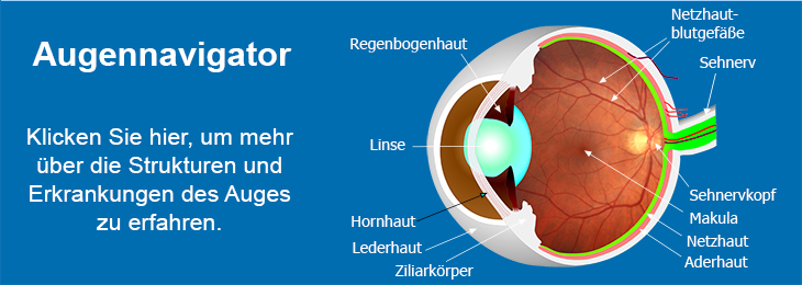
Age-related macular degeneration (AMD) is the major cause of legal blindness in the Western world. The prevalence of the disease increases dramatically with increasing age. In the European Eye Study, half of the 5000 participants being 65 years or older displayed soft, distinct drusen or pigmentary abnormalities, more prominent fundus changes or signs of AMD. It is estimated that 4.5 million people are affected by AMD.
The initial stage of AMD appears to be an extensive formation of drusen, which are small circular deposits of insoluble protein and lipid aggregates containing, among others, amyloid betapeptide, advanced glycation end products, apolipoprotein B & E, various complement factors, peroxidised lipids, MHC class II antigens, vitronectin and others. Moreover, lipofuscin accumulation in the retinal pigment epithelium (RPE) may play an important role in the pathogenesis of AMD, with N-retinylidene-N-retinylethanolamine (A2E) as a major fluorophore in RPE lipofuscin, as well as oxygen radicals, mutations and chronic inflammatory reactions.
As a consequence of these processes, the RPE starts to degenerate. This degeneration can be recognized as dark patchy areas in the macular region already described in 1977, and named “geographic atrophy” (GA) due to its appearance. In the areas where RPE cells die off, also photoreceptors and the choriocapillaris degenerate, which leads to a gradual progressive loss of function in the central visual field. In a number of patients, new blood vessels grow from the choroid into the subretinal space (choroidal neovascularisation, CNV). As the new vessels are not stable, they may leak. The resulting subretinal or sub-RPE haemorrhage can be visualised by fluorescein angiography and is the first sign of CNV in most cases. In order to distinguish these two major ways of AMD progression, GA and CNV, they are nominated “dry AMD” and “wet AMD”, respectively.
The microglia is the immune competent cell in the central nervous system (CNS), executing a variety of functions and showing different reactions to pathologies of the CNS. In the healthy eye, the subretinal space is immune privileged and devoid of microglia. However, microglial cells migrate into the subretinal space with increasing age or if photoreceptors degenerate. The same observation was made in AMD patients. Microglial cells activated by a disease release pro-inflammatory, chemotactic and pro-angiogenetic molecules, cause pathologic changes in the RPE and the blood-retina barrier and promote CNV.
RPE cells also attract other monocytes, which had been described already in 1987 without discrimination between the different kinds of monocytes. It became clear during the following years that macrophages and dendritic cells are involved in the pathologic processes occurring in AMD, e. g., in the local removal of Bruch’s membrane and the formation of drusen.
The goal of our research group is the investigation of the pathologic processes in the back of the eye connected with AMD. Moreover, we are interested in general issues of neovascularisation, also occurring in diabetic retinopathy, and interactions between the retina, RPE, choroid and the immune system.Our research aims especially at
- the processes of neovascularisation and properties of endothelial cells,
- the behaviour of the immune system, in particular the microglia, and its interaction with the RPE and blood vessels,
- the influence of lipofuscin and other pathologic factors on RPE cells and microglial cells in vitro and in vivo,
- the influence of polyphenols and other protective compounds on RPE cells, endothelial cells and microglial cells in vitro and in vivo, and
- the effects of gold nanoparticles (GNP) on RPE cells and endothelial cells, as well as utilisation of GNP in animal models of CNV.
As experimental models, we at first use in-vitro systems, i. e., pure and mixed cell cultures as well as retinal and choroidal explants. After various treatments, i. e., exposure to several compounds and conditions, changes in gene expression will be monitored, depending on the cell type, in particular of typical surface proteins, cytokines, growth factors and several enzymes. Moreover, behavioural parameters such as proliferation, phagocytosis and apoptosis will be monitored. For this purpose, the standard analytical techniques of RT-PCR, ELISA and fluorescence-activated cell sorting (FACS) will be applied.
We also intend to use various mouse strains as in vivo models, besides C57BL/6 mice also the mutant mouse strains CX3CR1+/-, CX3CR1-/-, CCR2-/-, CX3CR1-/-/CCR2-/- and LysM,Cre+. In these animals, features of AMD are developing with increasing age or after a laser-induced lesion of the RPE. Therefore, choroidal neovascularisation will be induced in a number of animals by a laser treatment. The choroidal neovascularisation and accompanying processes, such as microglial migration and proliferation, will be monitored in vivo by funduscopy, fluorescence angiography and optical coherence tomography (OCT). In addition, immunohistochemical analysis of the retina-choroid complex and FACS analysis of the cells in this region will be performed.
top


